Introduction
AI Lab Image Analysis Tools use artificial intelligence (AI), deep learning, computer vision, and image processing technologies to analyze scientific images generated from microscopes, medical imaging systems, laboratory instruments, and biological experiments.
Modern laboratories produce massive volumes of visual data from microscopy, histology, cell imaging, fluorescence imaging, and high-content screening workflows. Manually analyzing these images can be slow, inconsistent, and difficult to scale. AI-powered image analysis platforms help researchers automatically detect patterns, classify objects, measure biological features, and extract meaningful insights from complex images.
These platforms use technologies such as convolutional neural networks (CNNs), deep learning segmentation models, computer vision algorithms, and automated image interpretation workflows. They support applications including cell analysis, drug discovery, pathology research, neuroscience, genomics, and biotechnology.
AI Lab Image Analysis Tools integrate with microscopes, laboratory automation systems, image repositories, research databases, and scientific workflow platforms. They help researchers improve accuracy, accelerate experiments, and discover biological patterns while requiring expert scientific validation.
Real-world Use Cases
- Microscopy image analysis
- Cell segmentation and classification
- High-content screening
- Histology image analysis
- Drug discovery research
- Fluorescence imaging analysis
- Cellular phenotype detection
- Laboratory automation workflows
- Biological pattern recognition
- Scientific image quantification
Evaluation Criteria for Buyers
When selecting an AI Lab Image Analysis Tool, consider:
- AI image recognition capabilities
- Cell detection accuracy
- Segmentation performance
- Microscopy compatibility
- Automation capabilities
- Data visualization features
- Laboratory integration
- Scalability
- Security controls
- Research workflow support
Best For
- Biotechnology companies
- Pharmaceutical research teams
- Academic laboratories
- Clinical research organizations
- Life science researchers
Not Ideal For
Organizations expecting AI image analysis to replace scientific interpretation or laboratory validation.
Key Trends
- AI-powered microscopy
- Automated cell analysis
- Deep learning image segmentation
- High-content screening automation
- Digital pathology integration
- AI drug discovery workflows
- Computer vision in biology
- Cloud-based image analysis
- Automated laboratory research
- Multimodal biological analysis
Methodology
The platforms below were evaluated based on:
- AI image analysis capabilities
- Scientific workflow support
- Computer vision performance
- Integration capabilities
- Research adoption
- Scalability
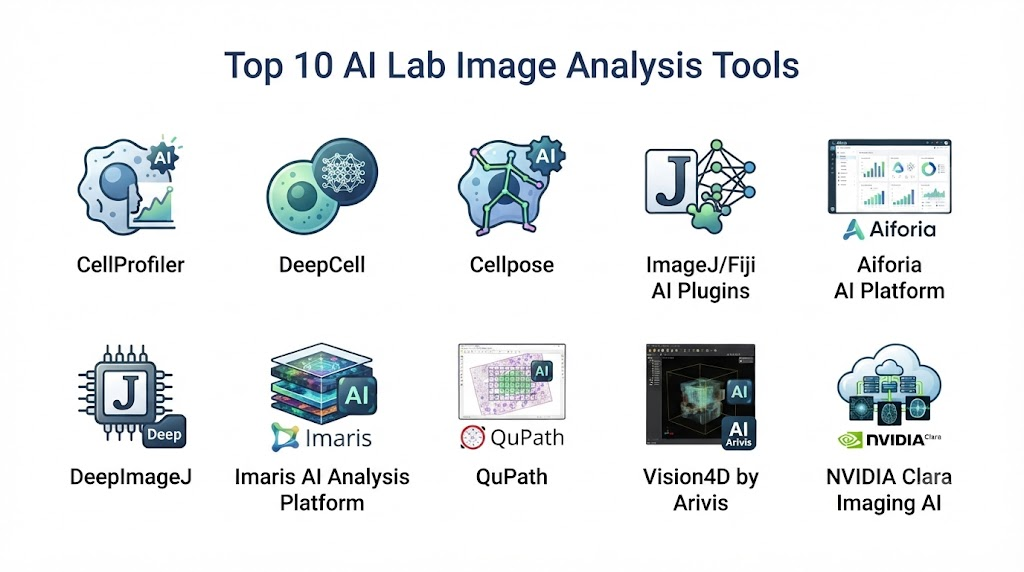
Top 10 AI Lab Image Analysis Tools
1. DeepCell
Verdict: Best overall AI-powered biological image analysis platform.
Short Description: DeepCell uses deep learning and computer vision technologies to analyze biological images, perform cell segmentation, and extract insights from microscopy data.
Key Features
- AI-powered cell segmentation
- Cell classification
- Microscopy analysis
- Image-based biological insights
- Automated workflows
Pros
- Advanced AI imaging capabilities
- Strong biological applications
- Reduces manual analysis effort
Cons
- Requires specialized imaging workflows
Deployment: Cloud and research environments
Security & Compliance: Research data protection controls
Integrations & Ecosystem: Microscopy systems, biological workflows, research platforms
Support & Community: Research and enterprise support
Pricing Model: Custom pricing
Best-Fit Scenarios: Cellular imaging research
2. CellProfiler
Verdict: Popular open-source platform for biological image analysis.
Short Description: CellProfiler provides automated image processing workflows for identifying and measuring biological objects in microscopy images.
Key Features
- Image processing pipelines
- Cell measurement
- Object detection
- Microscopy analysis
- Research automation
Pros
- Open-source flexibility
- Strong scientific adoption
Cons
- Requires workflow configuration
3. ImageJ / Fiji
Verdict: Widely used scientific image analysis ecosystem.
Short Description: ImageJ and Fiji provide powerful image processing and analysis capabilities used across research laboratories for microscopy and biological image analysis.
Key Features
- Image processing
- Scientific visualization
- Plugin ecosystem
- Quantitative analysis
- Microscopy support
Pros
- Large research community
- Highly customizable
Cons
- Requires technical expertise
4. NVIDIA Clara Imaging
Verdict: AI infrastructure platform for medical and scientific imaging workflows.
Short Description: NVIDIA Clara provides AI tools and computing infrastructure for developing advanced imaging applications using deep learning models.
Key Features
- AI image models
- Deep learning frameworks
- Image processing
- GPU acceleration
- Research development tools
Pros
- Powerful AI capabilities
- Supports large-scale imaging workloads
Cons
- Requires AI expertise
5. Zeiss ZEN AI Imaging Platform
Verdict: AI-supported microscopy analysis platform.
Short Description: ZEISS provides microscopy software solutions with AI capabilities for automated image analysis, segmentation, and scientific imaging workflows.
Key Features
- Microscopy image analysis
- AI segmentation
- Automated measurements
- Visualization tools
- Instrument integration
Pros
- Strong microscopy ecosystem
- High-quality imaging support
Cons
- Best suited for ZEISS workflows
6. Leica Microsystems AI Imaging Solutions
Verdict: Advanced microscopy platform with AI analysis capabilities.
Short Description: Leica provides microscopy and imaging solutions that incorporate AI-powered analysis for biological research and laboratory applications.
Key Features
- Microscopy automation
- AI image analysis
- Cell imaging
- Image processing
- Research workflows
Pros
- Strong laboratory integration
- High-quality imaging ecosystem
Cons
- Hardware-focused environment
7. QuPath
Verdict: Open-source platform for bioimage and pathology analysis.
Short Description: QuPath provides tools for analyzing large biological images, including microscopy and pathology datasets.
Key Features
- Image annotation
- Cell detection
- Tissue analysis
- Machine learning workflows
- Digital pathology support
Pros
- Open-source platform
- Strong research adoption
Cons
- Requires technical knowledge
8. Visiopharm AI Image Analysis Platform
Verdict: AI-powered image analysis solution for pathology and life sciences.
Short Description: Visiopharm uses artificial intelligence and image analysis algorithms to analyze tissue images and support biomedical research.
Key Features
- AI image analysis
- Tissue segmentation
- Biomarker analysis
- Quantitative pathology
- Research workflows
Pros
- Strong biomedical applications
- Advanced AI analysis
Cons
- Specialized platform
9. PerkinElmer Harmony High-Content Imaging
Verdict: High-content screening image analysis platform.
Short Description: PerkinElmer Harmony provides automated image analysis capabilities for cellular imaging, screening, and pharmaceutical research.
Key Features
- High-content screening
- Cell analysis
- Image classification
- Phenotypic analysis
- Research automation
Pros
- Strong drug discovery workflows
- Automated analysis capabilities
Cons
- Enterprise research focus
10. OpenAI-Based Custom AI Lab Image Analysis Assistant
Verdict: Flexible AI assistant for customized scientific image workflows.
Short Description: Research organizations can build custom AI lab image analysis assistants using computer vision models integrated with microscopy systems, image repositories, laboratory databases, and scientific workflows. These assistants can summarize image findings, organize analysis results, support annotations, and assist researchers while requiring expert validation.
Key Features
- Image interpretation support
- Research summaries
- Annotation assistance
- Workflow automation
- Scientific reporting
Pros
- Highly customizable
- Flexible integrations
- Improves researcher productivity
Cons
- Requires imaging expertise
- Validation required
Comparison Table
| Platform | AI Image Analysis | Microscopy Support | Automation | Research Integration | Best Use |
|---|---|---|---|---|---|
| DeepCell | Excellent | Excellent | Excellent | High | Cellular Imaging |
| CellProfiler | High | Excellent | High | High | Biological Image Processing |
| ImageJ/Fiji | Medium | Excellent | Medium | High | Research Imaging |
| NVIDIA Clara | Excellent | High | Excellent | High | AI Imaging Development |
| ZEISS ZEN AI | High | Excellent | High | Excellent | Microscopy Workflows |
| Leica AI Imaging | High | Excellent | High | Excellent | Laboratory Imaging |
| QuPath | High | High | High | High | Tissue Analysis |
| Visiopharm | Excellent | High | Excellent | High | Digital Pathology |
| Harmony | High | Excellent | Excellent | High | High Content Screening |
| OpenAI Custom | Custom | Custom | Custom | Custom | AI Image Assistant |
Evaluation & Scoring Table
| Platform | AI Capability 20% | Image Accuracy 20% | Automation 15% | Integration 15% | Security 10% | Ease 10% | Value 10% | Total |
|---|---|---|---|---|---|---|---|---|
| DeepCell | 20 | 20 | 15 | 14 | 10 | 8 | 8 | 95 |
| NVIDIA Clara | 20 | 19 | 15 | 14 | 10 | 8 | 8 | 94 |
| Visiopharm | 19 | 19 | 15 | 14 | 10 | 8 | 8 | 93 |
| ZEISS ZEN AI | 18 | 19 | 14 | 15 | 10 | 8 | 8 | 92 |
| Leica AI Imaging | 18 | 18 | 14 | 15 | 10 | 8 | 8 | 91 |
| Harmony | 18 | 18 | 15 | 14 | 10 | 8 | 8 | 91 |
| QuPath | 17 | 18 | 14 | 13 | 10 | 9 | 9 | 90 |
| CellProfiler | 17 | 18 | 13 | 14 | 10 | 9 | 9 | 90 |
| ImageJ/Fiji | 16 | 17 | 12 | 14 | 10 | 9 | 9 | 87 |
| OpenAI Custom | 20 | 16 | 12 | 15 | 8 | 7 | 9 | 87 |
Which AI Lab Image Analysis Tool Is Right for You?
| If your priority is… | Recommended Platform |
|---|---|
| AI cellular image analysis | DeepCell |
| AI imaging infrastructure | NVIDIA Clara |
| Open-source biological imaging | CellProfiler |
| Scientific image processing | ImageJ/Fiji |
| Microscopy automation | ZEISS ZEN AI |
| Laboratory microscopy workflows | Leica AI Imaging |
| Tissue image analysis | QuPath |
| Digital pathology research | Visiopharm |
| High-content screening | Harmony |
| Custom AI image assistant | OpenAI-Based AI Assistant |
Implementation Playbook
First 30 Days
- Define imaging analysis goals
- Identify image data sources
- Review microscopy workflows
- Select AI requirements
Days 31–60
- Configure image analysis pipelines
- Integrate imaging systems
- Train research teams
- Validate AI results
Days 61–90
- Automate image workflows
- Improve analysis accuracy
- Integrate research databases
- Expand AI-powered experiments
Common Mistakes
- Using poor-quality images
- Ignoring image preprocessing
- Overestimating AI accuracy
- Lack of scientific validation
- Poor workflow integration
- Ignoring data security
- Choosing unsuitable models
- Not involving domain experts
Frequently Asked Questions
1. What are AI Lab Image Analysis Tools?
They are AI-powered platforms that analyze scientific images and extract biological insights.
2. How does AI improve laboratory image analysis?
AI helps detect patterns, classify objects, segment cells, and automate image interpretation.
3. Can AI replace laboratory scientists?
No. AI supports researchers but requires expert review and validation.
4. Who uses AI image analysis tools?
Biotechnology companies, pharmaceutical researchers, academic laboratories, and healthcare organizations.
5. What images can these tools analyze?
They analyze microscopy images, cellular images, tissue images, and scientific imaging data.
6. Can AI improve drug discovery?
Yes. AI image analysis supports screening, cell analysis, and biological research.
7. Are AI image analysis results accurate?
Accuracy depends on image quality, model performance, and validation methods.
8. Do these tools integrate with microscopes?
Many platforms integrate with laboratory imaging systems and scientific instruments.
9. How is image data protected?
Organizations should use secure storage, access controls, and research data governance.
10. What should buyers evaluate before adoption?
Consider AI capabilities, imaging compatibility, integrations, scalability, security, and workflow needs.
Conclusion
AI Lab Image Analysis Tools are transforming scientific research by enabling faster, more accurate, and automated interpretation of complex laboratory images. By combining artificial intelligence, computer vision, and biological data analysis, these platforms help researchers discover cellular patterns, improve experiments, and accelerate innovation.Organizations adopting AI image analysis solutions should focus on image quality, scientific validation, integration capabilities, and workflow requirements. Platforms such as DeepCell, NVIDIA Clara, CellProfiler, ZEISS ZEN AI, and Visiopharm demonstrate how artificial intelligence is advancing laboratory imaging and supporting the future of biotechnology research.
