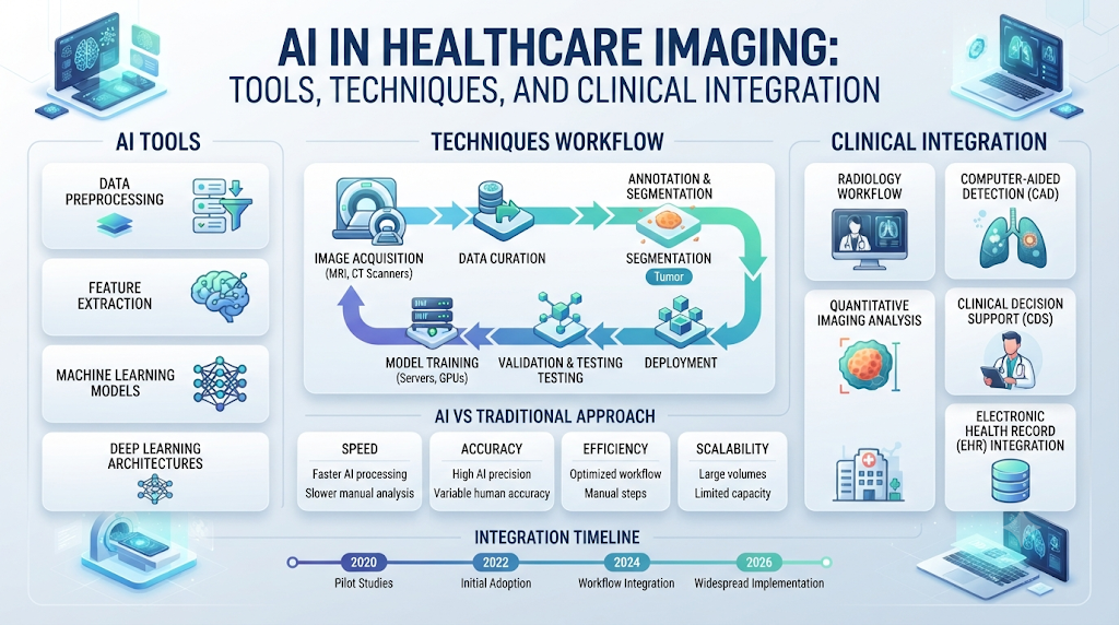
Medical imaging produces petabytes of data annually, ranging from routine chest radiographs and cross-sectional computed tomography (CT) to high-resolution magnetic resonance imaging (MRI) and digital pathology slides. At the same time, health systems face mounting workforce shortages and rising imaging volumes. Artificial intelligence (AI) and machine learning (ML) have shifted from experimental research into standard clinical infrastructure, functioning as critical decision-support layers across radiology, cardiology, and pathology.
Deploying AI in healthcare imaging requires understanding how these tools actually function within hospital networks, distinguishing between detection, triage, and automated reporting systems, and navigating rigorous validation and regulatory frameworks.
The Functional Landscape: Detection, Triage, and Reporting
“AI in healthcare imaging” is not a single technology category. Software applications deployed in clinical practice perform distinct operational jobs, operate under specific regulatory clearances, and interact with the Picture Archiving and Communication System (PACS) in different ways.
[Modality: CT / MRI / X-Ray]
       │
       ▼
[PACS / RIS Infrastructure]
       │
       ├─► [Triage & Notification] ──► Flags urgent studies (e.g., LVO Stroke, PE) on worklist
       ├─► [Detection & Analysis] ──► Highlights findings (e.g., nodules, fractures, hemorrhage)
       └─► [Generative Drafting]  ──► Summarizes findings into structured report templates
1. Narrow Detection and Classification Tools
These algorithms are trained to recognize specific, predefined abnormalities on a single modality. Examples include identifying pulmonary nodules on chest CT, subtle scaphoid fractures on plain radiographs, or intracranial hemorrhage on non-contrast head CT. They typically output bounding boxes, probability scores, or segmentation overlays that highlight regions of interest for the reading physician.
2. Workflow Triage and Prioritization Tools
Triage software does not provide a diagnostic interpretation. Instead, it runs silently in the background as studies enter the PACS, evaluating DICOM images for time-sensitive emergency conditions such as large-vessel occlusion (LVO) stroke, massive pulmonary embolism, or tension pneumothorax. If an acute finding is suspected, the system reorders the radiologist’s worklist or triggers a direct mobile alert to the care team, significantly reducing door-to-treatment times.
3. Quantitative Imaging and Biomarkers
AI models excel at automated measurement tasks that are tedious or subjective when performed manually. This includes volumetric analysis of liver lesions or brain tumors, precise quantification of coronary artery calcium and plaque burden, and automated calculation of skeletal bone age or Cobb angles in scoliosis assessment.
4. Generative Drafting and Structured Reporting
Natural language processing (NLP) and generative models assist radiologists by summarizing patient history, extracting prior imaging comparisons, and drafting structured report templates. When integrated correctly, these tools minimize documentation fatigue, though the final clinical interpretation and sign-off remain strictly under human physician control.
Core Techniques and Underlying Mechanics
Most commercial medical imaging AI relies on supervised deep learning, specifically convolutional neural networks (CNNs) and transformer-based vision models, trained on large annotated datasets of medical images.
- Image Reconstruction and Enhancement: Deep learning models reconstruct high-fidelity diagnostic images from sparse or low-dose raw data. In computed tomography and magnetic resonance imaging, these techniques reduce acquisition scan times or lower radiation doses without compromising diagnostic quality.
- Feature Extraction and Representation: Unlike human observers who look at global patterns, neural networks analyze pixel-level spatial hierarchies, detecting mathematical variations in texture, density, and boundary definition that may be imperceptible to the naked eye.
- Domain Adaptation and Cross-Vendor Generalization: A persistent engineering challenge is domain shift. An algorithm trained exclusively on scans from a 3-Tesla scanner made by one manufacturer may experience a drop in sensitivity or specificity when deployed on a 1.5-Tesla machine from a different vendor. Robust clinical pipelines require rigorous multi-center validation to account for hardware variance and patient demographics.
Decision Guidance: Evaluating and Procuring Imaging AI
Implementing AI in a clinical practice requires evaluating software through a structured operational and clinical framework.
| Evaluation Dimension | Key Considerations | What to Verify |
| Clinical Validation | Does the tool match your patient population? | Look for prospective, multi-site validation studies rather than retrospective internal datasets alone. |
| Regulatory Clearance | What is the exact intended use? | Check FDA 510(k), De Novo clearances, or CE mark indications to ensure the tool is legally cleared for your specific diagnostic workflow. |
| Workflow Integration | How does it deliver results to the clinician? | Seamless DICOM/PACS integration with minimal click fatigue; alerts must reach the right user without causing notification fatigue. |
| Total Cost of Ownership | Per-study licensing vs. enterprise subscription | Factor in ongoing IT maintenance, DICOM routing hardware, validation overhead, and regular model drift monitoring. |
When to Implement AI Imaging Tools
- High-Volume Screening: High-throughput modalities like mammography or diabetic retinopathy screening where algorithms can reliably rule out normal studies or flag subtle lesions.
- Emergency Triage Environments: Stroke or trauma centers where every minute saved in worklist prioritization directly impacts patient outcomes.
- Resource-Constrained Facilities: Outpatient centers or remote clinics lacking subspecialist coverage, provided the software acts as an assistive safety net.
When NOT to Use AI Imaging Tools
- As Autonomous Diagnostic Agents: Except for very narrow, specifically cleared screening applications (such as automated diabetic retinopathy screening in primary care), AI must not be used to issue final diagnostic reports without physician review.
- Out-of-Distribution Populations: If a model was validated primarily on adult urban cohorts, deploying it directly on pediatric or heavily underrepresented demographic populations without local performance verification risks high false-positive or false-negative rates.
Risks, Failure Modes, and Safety Governance
Deploying machine learning models in clinical environments introduces distinct failure modes that differ from traditional software bugs.
- Model Drift and Data Shift: Changes in hospital scanner hardware, contrast protocols, or patient demographics over time can degrade model accuracy. Continuous performance monitoring is necessary.
- Automation Bias: Clinicians may either over-rely on an AI algorithm’s output (accepting incorrect bounding boxes or missing genuine findings not flagged by the AI) or dismiss the tool entirely due to early false alarms.
- Adoption of Institutional Frameworks: Professional bodies, such as the American College of Radiology (ACR) and the Society for Imaging Informatics in Medicine (SIIM), emphasize establishing formal AI governance committees within health systems. These committees are responsible for vetting vendor claims, overseeing local validation, tracking algorithmic errors, and maintaining update lifecycles.
Frequently Asked Questions
Q1. Will AI replace radiologists in clinical practice? No. Essentially all clinically deployed radiology AI is designed to assist physicians rather than replace them. Regulatory bodies have not authorized any comprehensive diagnostic AI for autonomous, unreviewed final reporting. AI handles repetitive triage, measurement, and detection tasks, allowing physicians to focus on complex decision-making and patient communication.
Q2. What is the difference between diagnostic AI and triage AI?Diagnostic AI tools actively analyze images to detect, classify, or segment specific pathologies like nodules or fractures. Triage tools do not interpret disease; instead, they scan incoming studies for acute markers of critical conditions (such as intracranial hemorrhage or pulmonary embolism) and reprioritize the worklist so reading physicians examine urgent cases first.
Q3. How accurate are AI tools in medical imaging? Accuracy varies widely depending on the specific modality, target finding, and vendor. Published clinical trials often demonstrate that AI-assisted reading improves overall sensitivity for specific findings compared to unassisted reading. However, real-world performance can drop if the local scanner parameters or patient demographics differ from the manufacturer’s training dataset.
Q4. Are medical imaging AI tools approved by regulatory agencies? Yes. A large volume of software-as-a-medical-device (SaMD) applications have received FDA 510(k) clearances or CE marks under EU MDR guidelines. Most of these clearances use the 510(k) pathway, meaning they demonstrate substantial equivalence to existing predicate devices rather than undergoing de novo clinical trials.
Q5. How do AI algorithms integrate into hospital IT systems? AI tools typically integrate directly with the Picture Archiving and Communication System (PACS) and Radiology Information System (RIS) via DICOM communication standards. Studies route through an inference server where the AI processes the images and injects structured overlays, measurements, or worklist tags back into the standard viewing environment.
Q6. What are the primary limitations of current medical imaging AI? Key limitations include vulnerability to domain shift (poor performance on data from different scanner models), lack of contextual clinical reasoning outside the image pixels, potential bias from non-representative training data, and the overhead required to maintain continuous monitoring for model drift.
Q7. How do health systems pay for radiology AI software? Pricing models vary among vendors, including per-study fees, enterprise site licenses, or bundled software-as-a-service (SaaS) subscriptions. While adoption was initially driven by upfront capital investments in academic centers, increasing commercial insurance and public payer coverage for specific AI-analyzed diagnostics is expanding reimbursement pathways.
Q8. What is “model drift” in healthcare AI? Model drift occurs when the statistical properties of the incoming clinical data change over time—due to hardware upgrades, protocol shifts, or shifting patient populations—causing the AI algorithm’s diagnostic accuracy to degrade silently if left unmonitored.
Q9. Do hospitals need a dedicated AI governance committee? Yes. Professional guidelines recommend that healthcare facilities establish formal AI governance structures. These multidisciplinary teams consisting of radiologists, IT administrators, compliance officers, and physicists evaluate, test, deploy, and audit AI tools to ensure patient safety and data security.
Q10. What steps should a department take before purchasing an AI imaging tool? Facilities should clearly define the clinical problem they intend to solve, verify independent and prospective validation data, test integration compatibility with existing PACS architecture, review regulatory clearance documents, and establish local protocols for human-in-the-loop oversight.
Conclusion
Artificial intelligence in healthcare imaging provides powerful capabilities to streamline workflows, accelerate acute triage, and reduce diagnostic oversight. Success depends on rigorous clinical validation, thoughtful PACS integration, and structured institutional governance that treats AI as an assistive instrument rather than a standalone substitute for clinical judgment.